Performed by Dr. Praneeth Putti at Sharp Vision Eye Hospital
Retinal surgeries in Hyderabad address critical conditions affecting the retina, including retinal detachment, macular holes, diabetic retinopathy complications, and epiretinal membranes. These advanced microsurgical procedures are essential for preserving and restoring vision in patients with serious retinal disorders. At Sharp Vision Eye Hospital, Dr. Praneeth Putti combines cutting-edge surgical techniques with personalized care to deliver optimal outcomes for complex retinal conditions.
We believe in honest, upfront disclosure so you can make an informed decision.
A step-by-step look at what happens during this procedure.
The eye is numbed with local anaesthetic drops or injections, or general anaesthesia is administered depending on the complexity and patient preference.
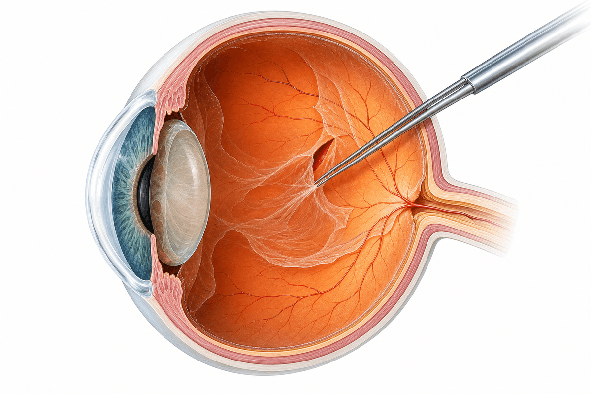
Tiny instruments are inserted through small incisions to remove the vitreous gel, allowing access to the retina and removing any blood, scar tissue, or debris.
The surgeon repairs retinal tears, reattaches detached retina using laser photocoagulation or cryotherapy, removes membranes, or closes macular holes as needed.
Gas bubble, silicone oil, or air is injected into the eye to hold the retina in place during healing, maintaining proper positioning for successful reattachment.
The tiny surgical incisions are self-sealing or closed with fine sutures, and antibiotic drops are applied to prevent infection.
A structured, patient-first approach from first consultation to full recovery.
Sharp Vision Eye Hospital utilizes state-of-the-art OCT, fundus photography, and ultrasound imaging to precisely map retinal pathology and plan the optimal surgical approach.
Our operating theaters are equipped with high-magnification microscopes and micro-instrumentation enabling Dr. Praneeth Putti to perform delicate retinal repairs with exceptional accuracy.
We provide detailed positioning instructions, customized medication regimens, and scheduled follow-up appointments to monitor healing and ensure optimal retinal reattachment.
Our team ensures patients understand their specific condition, surgical procedure, recovery expectations, and warning signs that require immediate attention throughout the treatment journey.
What to expect at each stage of your recovery journey.
Strict head positioning may be required if gas bubble was used. Vision will be blurry, and eye may feel uncomfortable. Avoid strenuous activities, heavy lifting, and follow prescribed eye drop regimen diligently.
Gradual vision improvement begins as the retina heals and gas bubble dissipates. Continue protective measures, attend all follow-up appointments, and report any sudden vision changes immediately.
Vision continues to improve as retinal function stabilizes. Most patients return to normal activities with ophthalmologist clearance. Final visual acuity depends on pre-operative damage and surgical success.
Over 90% of patients achieve successful anatomical retinal reattachment after primary surgery, preventing further vision loss and preserving remaining visual function.
Many patients experience significant vision improvement, though final visual acuity depends on duration and severity of detachment, with earlier treatment yielding better outcomes.
Timely retinal surgery prevents permanent vision loss and blindness from progressive retinal detachment, macular holes, or severe retinopathy complications.
Successful retinal surgery enables patients to resume daily activities, maintain independence, and experience improved confidence with restored or preserved vision.
Let's clear up some of the most common myths about retinal surgeries.
Get a personalised assessment from an experienced specialist. Understand your options, ask your questions, and decide with confidence.